ArchivesCategories |
Back to Blog
 Magnetic Resonance Imaging (MRI) Anatomical and physiological images of the body may be created using the medical imaging technology known as magnetic resonance imaging (MRI). To create pictures of the body's organs, MRI scanners use radio waves, intense magnetic fields, and magnetic field gradients.
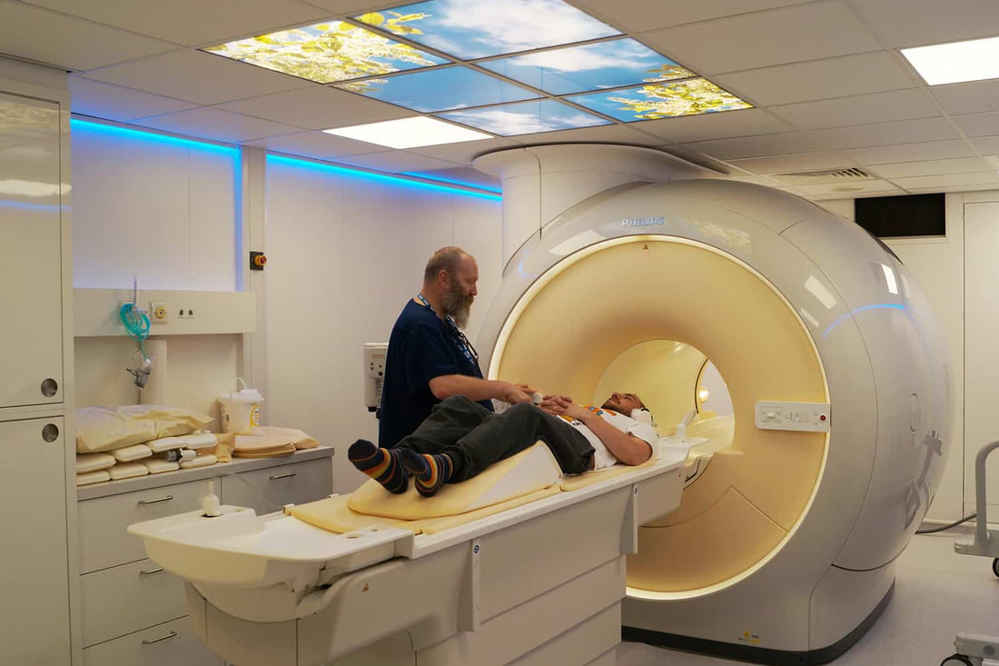
How does Magnetic Resonance Imaging (MRI) work? Powerful magnets are used in Magnetic Resonance Imaging (MRI) to create a strong magnetic field that compels the body's protons to align with it. After that, radiofrequency radiation is pumped into the patient, stimulating the protons and causing them to spin out of balance and battle the magnetic field. The MRI sensors can pick up the energy produced as the protons realign with the magnetic field when the radiofrequency field is switched off. Depending on the surroundings and chemical makeup of the molecules, both the time it takes for the protons to realign with the magnetic field and the quantity of energy released vary. These magnetic characteristics allow doctors to distinguish between different tissue kinds. MRI Used For A powerful magnet and radio waves are used in magnetic resonance imaging (MRI) to examine the organs and structures within your body. MRI scans are used by medical practitioners to identify a range of illnesses, from cancers to damaged ligaments. The brain and spinal cord may be examined with MRIs. Is an MRI safe? When necessary safety precautions are taken, an Magnetic Resonance Imaging (MRI) scan is typically safe and offers nearly no danger to the ordinary individual. The intense magnetic field produced by MRI scanners does not damage you, but it might interfere with implanted medical equipment or alter the pictures. If your MRI involves the use of contrast material, there is a very small chance that you will experience an allergic response. Often, these responses are minor and manageable with medicine. A medical professional will be on hand to give prompt aid if you experience an allergic reaction. Due to the unknown hazards to the unborn child, medical professionals often avoid doing gadolinium contrast-enhanced MRIs on pregnant patients unless it is absolutely required. What does the equipment look like? The conventional MRI machine is a huge tube with a cylinder form that resembles a gigantic doughnut. On a table that glides into a tube leading to the middle of the MRI machine, you will lay down. Those who suffer from acute claustrophobia may benefit from MRI units that are more "open" in form. For some tests, an open MRI is not a viable option. Consult your radiologist for further details. For bigger individuals or those who experience claustrophobia, several of the latest MRI scanners offer a wider diameter bore. Which is better a CT scan or MRI? Compared to a CT scan, magnetic resonance imaging gives pictures that are more clear. An MRI is preferable to X-rays or CTs when a view of soft tissues is required for medical professionals. Compared to CT images, MRIs can produce images of the organs and soft tissues that, for example, damaged ligaments and herniated discs.
0 Comments
Leave a Reply. |
 RSS Feed
