ArchivesCategories |
Back to Blog
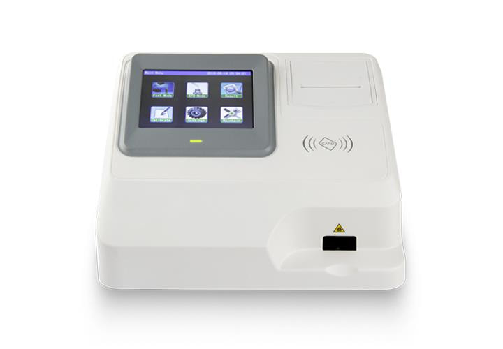 Immunofluorescence Analyzer The detection and measurement of certain proteins or antibodies in biological samples is made possible by the use of an immunofluorescence analyzer, a potent diagnostic tool utilised in clinical laboratories and medical research. For the purpose of locating and quantifying the presence of certain biomolecules inside cells or tissues, it combines immunology and fluorescence microscopy methods.
An immunofluorescence analyzer works by using fluorescently-labeled antibodies that attach to the target molecules selectively. Animals that have been exposed to the target antigens and the resulting immunological response are used to manufacture these antibodies. Following their purification, the antibodies are coupled with fluorescent dyes like FITC or rhodamine. The fluorescently-labeled antibodies are then treated with the biological material to be examined after it has been fixed onto a microscope slide. Any extra antibodies are washed away once the antibodies attach directly to the target molecules. A fluorescence microscope is used to see the material after it has been excited by light of a particular wavelength to reveal the fluorescent dye on the attached antibodies. As a result of the dye's longer wavelength light emission, the target molecules' position and intensity may be seen in an image produced by the microscope. Numerous medical illnesses, such as cancer, autoimmune diseases, and infectious diseases, are studied and diagnosed using immunofluorescence analyzers. An immunofluorescence analyzer, for instance, may find and measure the presence of viral antigens in biological samples like blood, urine, or sputum for diagnosing infectious disorders like viral infections. Utilising certain antibodies that bind to viral antigens enables the identification and measurement of the virus in the sample. Similar to this, an immunofluorescence analyzer may identify and measure the presence of autoantibodies that attack the body's own tissues in the diagnosis of autoimmune illnesses like rheumatoid arthritis. This makes it possible to identify and diagnose the illness early, which can help in the creation of effective treatment strategies. The expression of certain proteins or biomarkers linked to the onset and course of cancer can be found and quantified using immunofluorescence analyzers in cancer research. This can help with both the diagnosis and stage of the illness as well as the creation of specialised treatments. The excellent sensitivity and specificity of immunofluorescence analyzers make them an important tool for clinical diagnosis and medical research. To provide precise and repeatable findings, they call for meticulous sample preparation and handling. The accuracy and dependability of the results can be impacted by a number of variables, including the quality and specificity of the utilised antibodies, the incubation period, and the microscope settings. An immunofluorescence analyzer is a powerful diagnostic tool that combines immunology and fluorescence microscopy techniques to detect and quantify specific proteins or antibodies in biological samples. Its wide range of applications in medical research and clinical diagnosis makes it an invaluable tool for advancing our understanding of various diseases and developing targeted treatments. However, careful sample preparation and handling are essential to ensure accurate and reliable results.
0 Comments
Leave a Reply. |
 RSS Feed
